Enhancing Medical Consultation with Human Anatomical Models
Share
In the evolving landscape of healthcare, effective communication between doctors and patients is more important than ever. One tool that has stood the test of time in improving doctor-patient interactions is the use of human anatomical models. These 3D representations of human organs, bones, and systems are invaluable during consultations, helping doctors explain medical conditions, procedures, and treatment options with clarity and precision. Here’s a closer look at how these models benefit both doctors and patients.
Visualizing Complex Anatomy
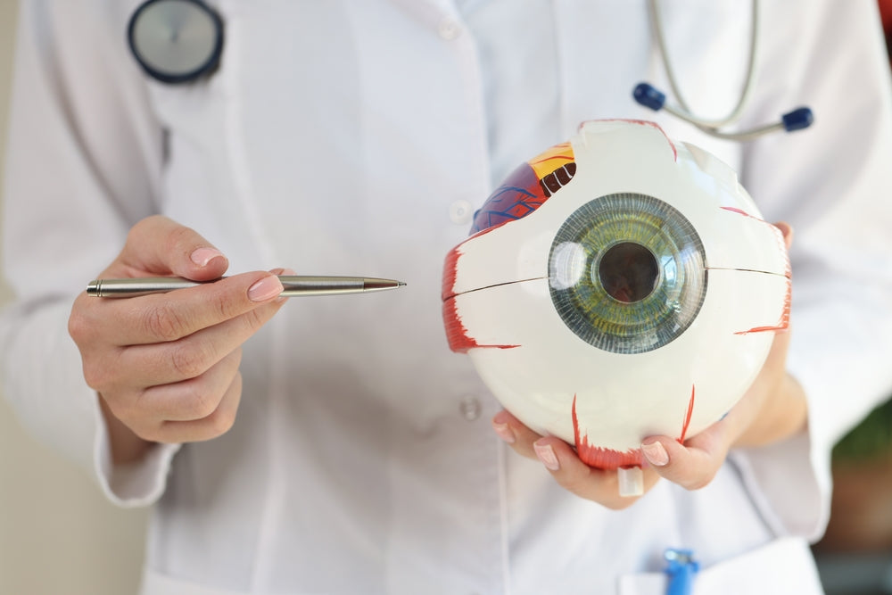
Human anatomy is intricate, and explaining its complexities verbally can be challenging. Anatomical models offer a tangible, visual representation of the body, making it easier for patients to grasp what’s happening inside them. Whether it’s a replica of a heart showing the chambers and blood flow or a model of the skeletal system highlighting joint function, these models break down complex ideas into something visible and understandable.
For instance, explaining heart disease or the need for surgery becomes far more effective when a doctor can point directly to a model of the heart, showing patients exactly where a blockage or problem lies. This visualization fosters better understanding, reducing anxiety and confusion.
Improving Patient Engagement
Anatomical models actively engage patients in their healthcare. Patients often feel overwhelmed by medical jargon, and it can be difficult to fully comprehend diagnoses or procedures. Models create an interactive consultation where patients can ask questions, point to areas of concern, and gain deeper insights into their condition. This active involvement leads to more informed decisions, increased satisfaction, and greater trust between doctor and patient.
For example, orthopedic doctors frequently use skeletal models to explain fractures or joint problems, enabling patients to see how their bones are affected and understand treatment options, whether that involves surgery or physical therapy.
Enhancing Communication Across Barriers
In today’s multicultural society, language barriers can present challenges in medical consultations. Even with translation services, medical terminology can be difficult to explain across languages. Anatomical models serve as a universal language, allowing doctors to communicate more effectively with patients who may not fully understand verbal explanations. This helps ensure that all patients, regardless of linguistic or educational background, have a clear understanding of their health and treatment.
Educational Value in Medical Training
Anatomical models are also key to the education and training of medical professionals. From medical students to experienced doctors, these models provide a hands-on learning experience that enhances theoretical knowledge. By practicing on 3D models, future doctors develop a better sense of spatial relationships within the body, improving their diagnostic and surgical skills. For seasoned professionals, models can refresh their understanding of specific anatomy, especially when dealing with rare or complex cases.
Building Trust and Reducing Anxiety
When patients understand their condition better, they are more likely to trust their doctor’s advice and feel confident about the proposed treatment. Anatomical models help bridge the gap between doctor and patient, promoting transparency in care. Being able to see exactly what’s wrong and how it can be fixed reduces the uncertainty and fear that often accompanies medical diagnoses.
For example, during consultations about surgical procedures, showing patients a model of the area being operated on demystifies the process. They can see how the surgery will be performed, which tissues will be affected, and what the outcome will look like. This significantly alleviates the fear of the unknown.
Incorporating Technology with Anatomical Models
While physical models have been the mainstay, technological advancements are enhancing their use. Digital anatomical models, including augmented reality (AR) and virtual reality (VR) versions, are gaining traction in modern medicine. These tools allow for interactive simulations where patients and doctors can view a 3D model of their own anatomy based on medical imaging, offering an even more personalized consultation experience.
For instance, AR can project a 3D model of a patient's heart onto a tablet, allowing the doctor to manipulate it in real-time, showing the patient dynamic aspects of their condition like blood flow or tissue abnormalities.
Cost-Effectiveness and Reusability
One of the benefits of physical anatomical models is their durability and reusability. Once purchased, a high-quality model can be used for years in various consultations, offering a cost-effective solution for clinics and hospitals. Compared to digital solutions, which may require software updates or subscriptions, physical models are a long-term investment that delivers consistent results.
Conclusion
Human anatomical models are an indispensable tool in medical consultations. They break down complex information into understandable visuals, engage patients in their own care, and enhance communication across barriers. As technology advances, integrating traditional models with digital solutions will only further improve patient outcomes and medical education. For doctors, these models are more than just teaching aids—they are a bridge to better patient care, deeper trust, and more successful treatments.
